
Double Bond Localization in Lipid Mass Spectrometry
In lipid structural analysis, the position of a carbon-carbon double bond within a fatty acid chain is a defining structural feature—not just its presence. Lipids with identical molecular formulas and the same number of double bonds can differ substantially in their biological behavior depending on exactly where those double bonds sit along the chain. This positional information is central to understanding structure-function relationships in lipidomics, nutrition science, and clinical lipid research.
Where Conventional Mass Spectrometry Falls Short
Standard mass spectrometry workflows are highly effective at determining a lipid’s molecular formula, carbon chain length, and overall degree of saturation. What they typically cannot resolve directly is the exact position of each double bond within the chain—because conventional fragmentation does not reliably break the molecule at that specific site.
To close this gap, two established approaches are widely used:
- Chemical derivatization, such as Paternò-Büchi reactions, which chemically tag the double bond prior to analysis
- External ozonolysis (OzID), which cleaves double bonds using ozone generated outside the ion source
Both methods are effective, but they share the same drawback: they add a dedicated reagent, an additional reaction step, and increased method complexity—along with the variability that extra sample handling introduces.
The Mechanism: In-Source Oxidative Cleavage via DBDI
Dielectric Barrier Discharge Ionization (DBDI), as implemented in SICRIT® technology, is a gas-phase, ambient ionization technique, sitting alongside familiar approaches such as ESI, APCI, and APPI. What sets it apart is a mechanistic side effect: the radical-driven processes underlying DBD ionization also generate reactive oxygen species—effectively producing ozone and peroxide intermediates in situ.
This means ionization and controlled oxidative cleavage at the double bond can occur within a series of radical reactions, directly in the ion source, at the MS1 level—with no added dopants and no separate derivatization step.
The key controlling variable is solvent composition. With an organic, nonpolar solvent gradient, lipid species ionize normally and no cleavage is observed. Switching to an aqueous, polar solvent system activates the oxidative mechanism, producing controlled, double-bond-specific cleavage for each unsaturation in the chain. This shifts double-bond localization from a separate analytical step to a property of the solvent system within an otherwise standard LC-MS or direct-infusion workflow.
What the Method Delivers
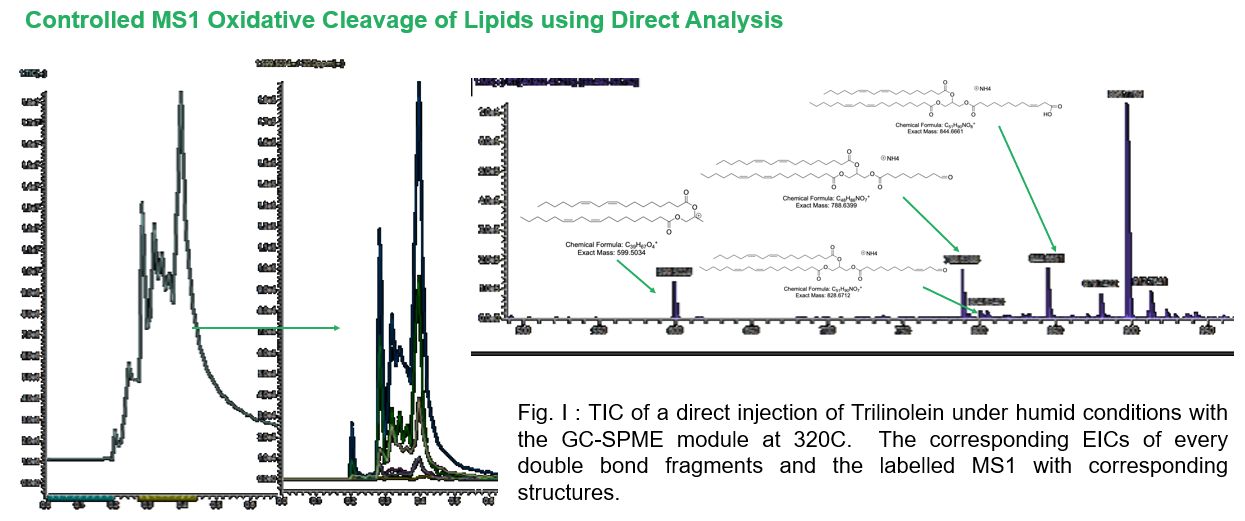
This aqueous DBDI approach has been shown to produce consistent, double-bond-specific oxidative fragments across a range of lipid classes, including triacylglycerols (TAGs) of varying saturation, as well as more polar lipid classes such as phosphatidylethanolamines (PE), phosphatidylcholines (PC), and cholesteryl esters (CholEster). The resulting fragments are reproducible and can be further confirmed at the MS2 level.
Notably, the method also works without chromatographic separation: direct analysis under humidified conditions alone is sufficient to generate the characteristic cleavage pattern—offering a faster alternative for applications where full LC separation isn’t required.
Outlook
Compared to dopant-assisted ozonolysis or derivatization-based methods, this DBDI-based approach removes the need for external dopants and additional method steps, while working within the ionization step researchers already rely on. Ongoing work focuses on further optimizing solvent and gas conditions to increase the yield of in-source oxidative radical species, with the goal of improving sensitivity and broadening applicability across lipid classes.
This mechanism builds on the broader capabilities of SICRIT® ionization technology, which supports a wide polarity range of analytes beyond lipid structural analysis.
This post was created with the assistance of AI and editorially reviewed.