MS
Imaging
Mass spectrometry (MS) imaging visualizes the spatial arrangement of molecules, such as biomarkers, metabolites, peptides, or proteins, based on their molecular masses. The SICRIT® Ion Source in combination with a laser ablation setup or as post-ionization device for (AP-) MALDI enables MS imaging down to 5μm.
The SICRIT® Ion Source in combination with a laser ablation setup or as post-ionization device for (AP-) MALDI enables MS imaging down to 5μm.
High Resolution Laser Ablation
The combination of a UV-laser ablation system with the flow-through SICRIT® Ion Source allows for a efficient, soft ionization-based visualization of molecules with a very high spatial resolution.
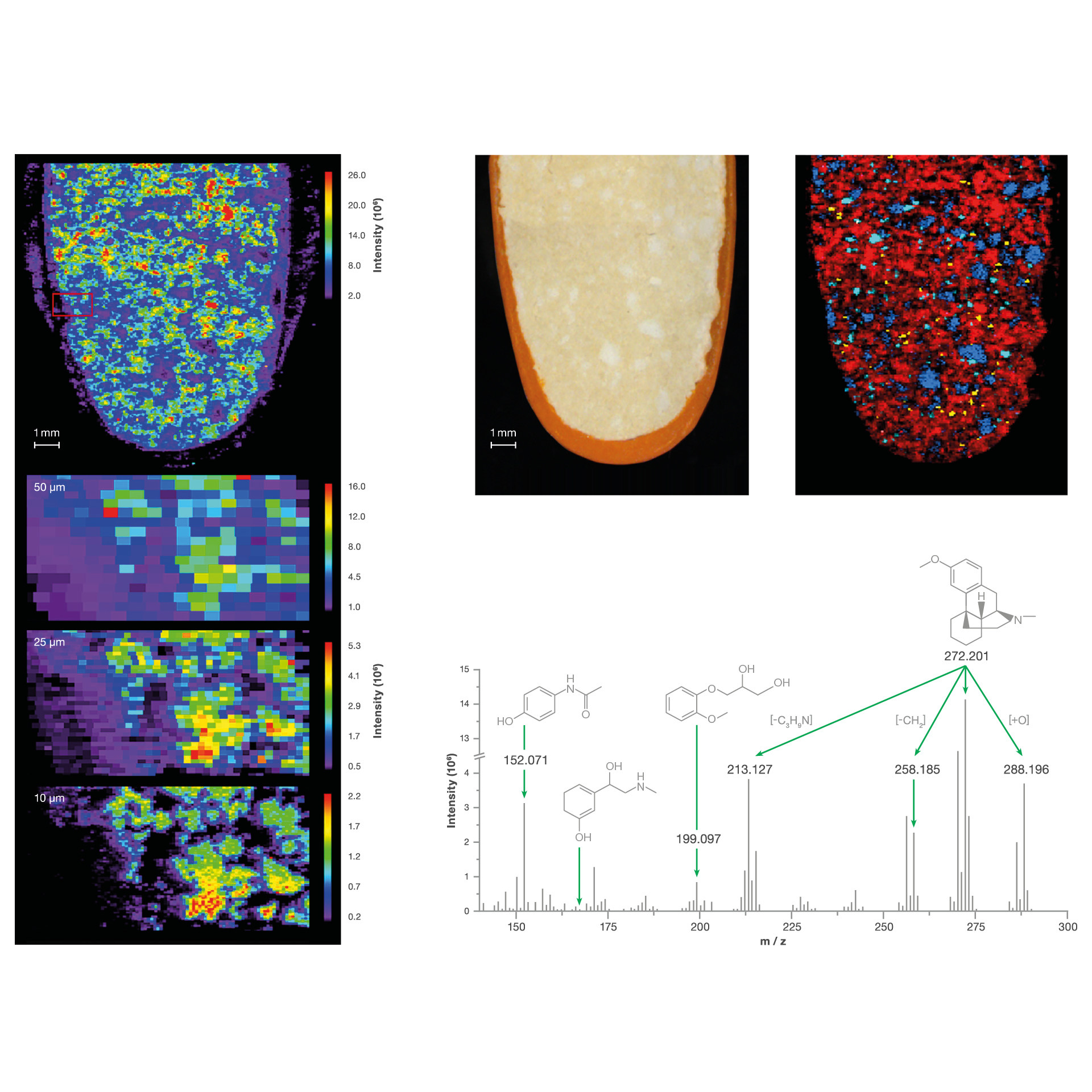
The SICRIT® Ion Source can be hyphenated to any commercial laser ablation system and nearly any API mass spectrometer that is commercially available. This makes this technique easy to adapt to individual needs regarding the sample type and analytical question.“
Elia, E.A. et al, Anal. Chem. 2020, 92, 15285–15290
Postionization For (AP)Maldi
Using the flow-through SICRIT® Ion Source as post-ionization device for AP-MALDI imaging allows to ionize and visualize additional molecules that have not been ionized with the primary ionization source. This significantly expands the mass spectrometric view on the sample in an MS imaging approach.
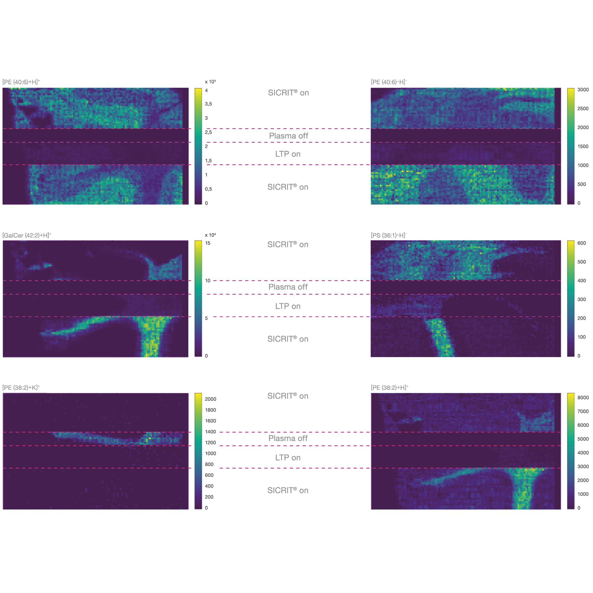
The ease with which the SICRIT® device can be installed and the minimal need for optimization presents this commercially available tool as an attractive method for simple post-ionization for any AP-MALDI MS imaging.“
Elia, E.A. et al, Anal. Chem. 2020, 92, 15285–15290
Knowledge Base
Subcellular MSI of lipids and nucleotides using transmission geometry ambient laser desorption and SICRIT® ionization
A plasma-assisted transmission-geometry laser desorption method enables subcellular MS imaging of lipids and nucleotides at 1 µm resolution, enhancing ionization efficiency and revealing spatial molecular distributions within tissues.
Discover our Products
Find the perfect solution for your use case, with Plasmion’s advanced technology and expertise in chemical analysis for research or industry.


