MS Imaging: Mapping Molecules with Precision
Mass Spectrometry Imaging is changing the way researchers explore the chemical composition of samples. By combining high-resolution mass spectrometry with spatial mapping, it creates a molecular picture of a surface. And with new approaches in ionization, MS Imaging is becoming even more powerful and versatile.
What is MS Imaging?
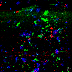
MS Imaging is a technique that merges two worlds: analytical chemistry and imaging. Instead of simply identifying which molecules are present in a sample, MS Imaging also shows their exact location. The result is a detailed chemical map that reveals how compounds are distributed across tissues, pharmaceutical tablets, food products, or materials.
Applications of MS Imaging
MS Imaging has become an essential tool across different scientific fields:
- Pharmaceutical research: Determining how active pharmaceutical ingredients (APIs) are distributed within a tablet or across tissue samples.
- Life sciences and medicine: Identifying biomarkers, monitoring disease progression, or tracking metabolic changes.
- Material sciences: Understanding chemical heterogeneity in polymers, coatings, or nanomaterials.
- Food and nutrition research: Mapping authenticity, additives, or contaminants in complex matrices.
Wherever location-specific chemical information is critical, MS Imaging techniques provide insights that traditional methods cannot.
How does an MS Imaging analysis work?
A typical MS Imaging workflow can be summarized in a few key steps:
- Sample preparation: The sample surface is mounted for analysis.
- Laser desorption: A finely focused laser shoots molecules off the surface, point by point.
- Ionization: The desorbed molecules are ionized, a crucial step in MS Imaging ionization.
- Mass Spectrometry Detection: The mass spectrometer measures the mass-to-charge ratio and abundance of ions for each sampled spot, generating raw spectra.
- Data Processing Software: Specialized software reconstructs the raw spectra into spatially resolved molecular images, showing the distribution of compounds across the sample surface.
This process delivers a molecular landscape, showing exactly where different compounds are located and in what relative abundance.


MS Imaging versus other imaging techniques
Every imaging method has its place. Microscopy, autoradiography, or fluorescence imaging can provide valuable structural or functional information, depending on the scientific question. MS Imaging adds a unique perspective: it directly measures the chemical composition of a surface without the need for labels or markers.
Thanks to MS Imaging, multiple analytes can be detected simultaneously, and even unexpected compounds can be discovered. This makes the technique especially valuable when unbiased, chemically specific information is required.
Ion sources for MS Imaging Ionization
The efficiency and scope of MS Imaging strongly depend on the ionization technique applied. Established ion sources such as MALDI (Matrix-Assisted Laser Desorption/Ionization), DESI (Desorption Electrospray Ionization), and SIMS (Secondary Ion Mass Spectrometry) have each proven valuable for different sample types and research questions.
Recently, new approaches have been developed to broaden the capabilities of MS Imaging ionization. One of these is SICRIT® Ionization Technology. This ion source applies a gentle ionization mechanism that is compatible with a wide range of molecules and existing lab setups. It can extend the range of detectable analytes, and simplify workflows, making it an attractive complement to established ion sources.
Learn more about MS Imaging in practice
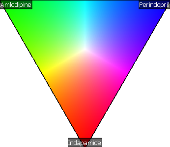
MS Imaging is more than just a method, it’s a way to visualize chemistry in space. A practical example is the analysis of active pharmaceutical ingredients on the surface of a tablet, where MS Imaging ionization reveals how compounds are distributed.
👉 Download our Application Note to see how this type of analysis was carried out and what insights it provided for pharmaceutical research.
